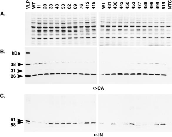
FIG. 4.
Immunoblot analysis of Ty3 proteins. (A) A 1-μg portion of wild-type Ty3 VLP protein or 10 μg of whole-cell extract isolated from yTM443 cells overexpressing wild-type Ty3 (WT) or IN mutant Ty3 or from nontransformed cells (NTC) was separated on a denaturing SDS–10% polyacrylamide gel and stained with Coomassie blue. (B and C) Identical samples were transferred to nitrocellulose (Hybond ECL; Amersham) and subjected to immunoblot analysis with a polyclonal rabbit anti-CA immunoglobulin G antibody (B) or a polyclonal rabbit anti-IN immunoglobulin G antibody (C). The positions of the structural proteins p38 (38 kDa), p31 (31 kDa), and CA (26 kDa) and the IN species (61 and 58 kDa [apparent only for VLP protein]) are indicated on the left. The 115-kDa RT-IN fusion protein was not detectable with anti-IN antibody on immunoblots of whole-cell extracts.

