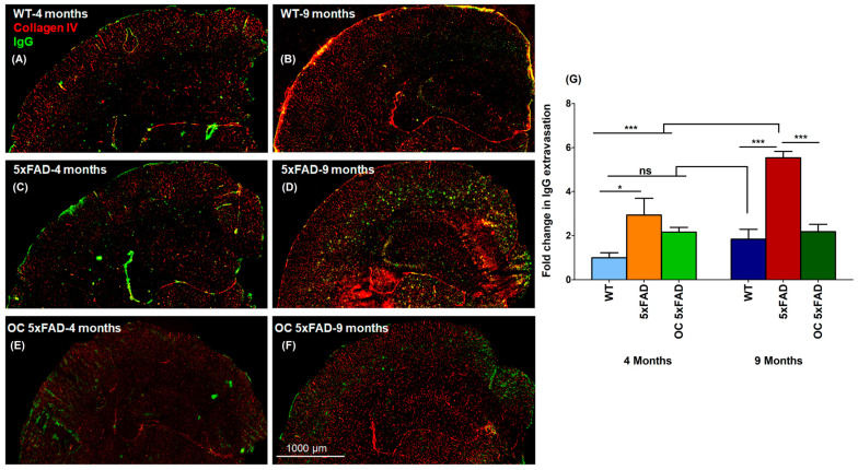
Figure 2.
Representative brain sections stained with anti-mouse IgG antibody to detect IgG extravasation (green) and anti-collagen antibody (red) in mouse brain (A) WT-4-month-old, (B) WT-9-month-old, (C) 5xFAD-4-month-old, (D) 5xFAD-9-month-old, (E) 5xFAD-4-month-old treated with 10 mg/kg OC daily for 3 months, and (F) 5xFAD-9-month-old treated with 10 mg/kg OC daily for 3 months. (G) IgG optical density in mice brains was quantified for IgG extravasation. Data are presented as mean + SEM for n = 5 mice/group. ns for not significant, * p < 0.05, and *** p < 0.001 vs. WT-4 months WT mice. Scale bar, 1000 μm.

