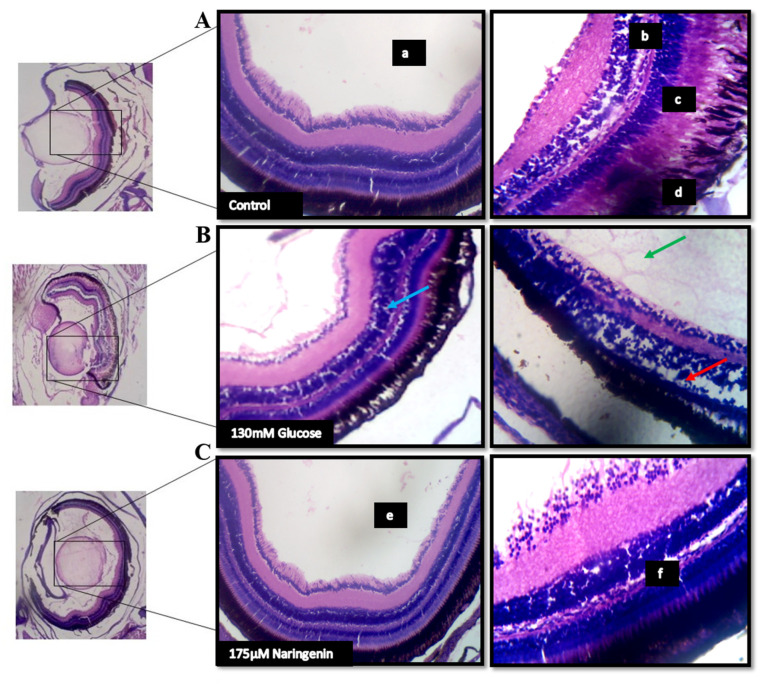
Figure 7.
The histopathological examination of zebrafish eye (Lateral section): (A) Control; (B) Model group (Glucose treated); (C) Treatment group; (a) lens; (b) retinal cells; (c) rods and cones; (d) Pigment epithelium in the control group. The red arrow indicates a disruption in the retinal cell layer; the blue arrow indicates the vacuole formation and necrosis in the rods and cones layer; the green arrow indicates the massive destruction in the lens. (e) lens and (f) the layers of the retina being normalized after the treatment.

