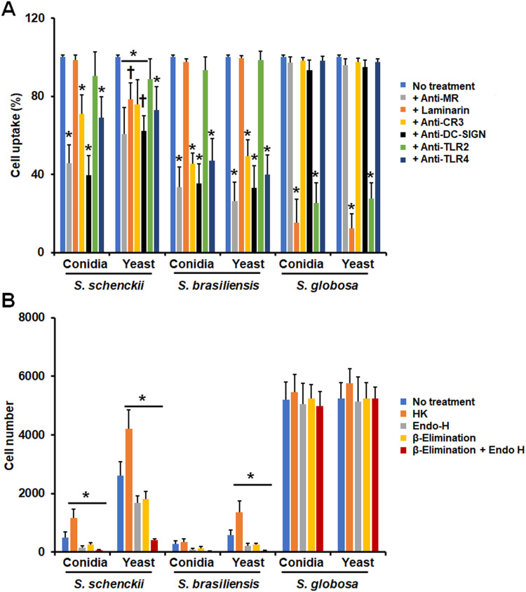
Figure 7.
Contribution of pattern recognition receptors and cell wall components on the phagocytosis of Sporothrix schenckii, Sporothrix brasiliensis, and Sporothrix globosa by human monocyte-derived Dendritic cells. In (A), Immune cells were preincubated with 200 μg mL−1 laminarin or 10 μg mL−1 of any of the following antibodies: anti-mannose receptor (MR), anti-CR3, anti-DCSIGN, anti-TLR2, or anti-TLR4. Then, cells were coincubated with Acridine Orange-labeled conidia or yeast-like cells at an immune cell-fungus ratio of 1:6, for 2 h at 37°C and 5% (v/v) CO2. Human cells were analyzed by flow cytometry, collecting 50,000 events, which were defined as a human cell interacting with at least one fluorescent fungal cell. All the interactions were performed in the presence of 5 μg mL−1 polymyxin B. No treatment refers to cells preincubated only with PBS. Results correspond to cells in the late stage of phagocytosis. For all cases, 100% corresponds to the system with no treatment, and the absolute values were similar to those shown in Figure 6 or (B) of this figure. CR3, complement receptor 3. *P < 0.05 when compared to the no-treatment condition of the same strain. †P< 0.05 when compared to conidia from the same species. In (B), Similar experiments as described in (A), but human cells were not preincubated with any blocking agent. Before coincubation with human monocyte-derived dendritic, conidia and yeast-like cells were inactivated by heat (HK) treated with endoglycosidase H (Endo-H), β-eliminated to remove O-linked glycans, or both treated with endoglycosidase H and β-elimination. No treatment refers to live cells without any treatment. *P < 0.05 when compared to the no-treatment condition of the same strain. Results are shown as mean ± standard deviation from data generated with samples from eight donors analyzed by duplicate.

