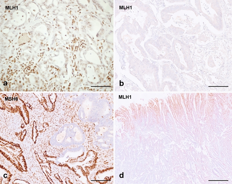
Fig. 2.
a MMR-deficient biopsy case showing complete loss of MLH1 expression in nuclei with minimal punctate nuclear expression—this type of expression is usually interpreted as loss of expression and leads to a diagnosis of deficient MMR. b Case of inadequate MMR immunostaining (surgical case), as no nuclear expression is seen in either neoplastic or internal control (stromal and inflammatory cells); this case was MSS by MSI testing. c Case of MMR-deficient tumour with loss of expression of MLH1 and PMS2 (not shown) and sub-clonal loss of MSH6 (surgical case). d pMMR colorectal cancer resection specimen showing central artefact with preserved expression in neoplastic and control cells towards the periphery, and loss of expression of both neoplastic and control cells in the central part of the tumour (probably due to hypo-fixation of the central area). Scale bar in a, b and c—50 µm, magnification ×40; scale bar in d—200 µm, magnification ×4

