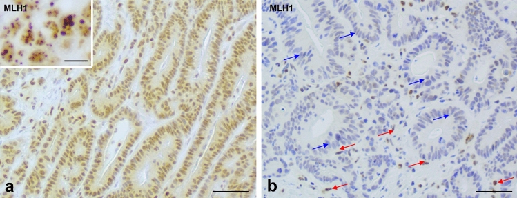
Fig. 4.
a Biopsy case from a MLH1/PMS2 MMR-deficient colorectal cancer showing MLH1 immunostaining with relatively weak nuclear expression and punctate nuclear expression (see inset). b This is the same case as a, subsequent to modification of immunohistochemistry protocol (protocol A) with reduced primary antibody incubation times. The red arrows show that the internal control cell nuclei have maintained expression while the neoplastic cells show greatly reduced spurious positivity (blue arrows). Scale bar—50 µm, magnification ×40 (10 µm in inset, magnification ×63)

