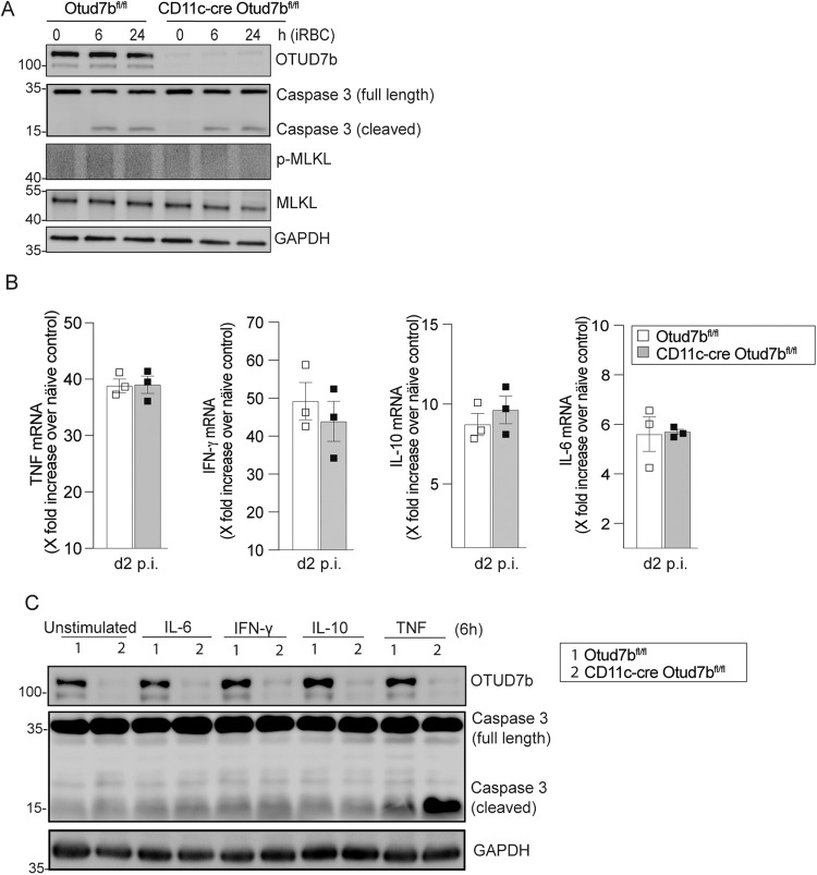
Fig. 2. OTUD7b confers protection against apoptosis upon TNF stimulation.
A Bone marrow-derived dendritic cells (BMDCs) were incubated with PbA-infected RBCs at a ratio of 1:3 (DC:iRBCs). Cells were harvested at indicated timepoints. Isolated proteins were analyzed by WB for the indicated proteins. Representative WB of individual mice are shown (n = 3 per group). B Otud7bfl/fl and CD11c-Cre Otud7bfl/fl mice were infected i.p. with 1 × 10⁶ iRBCs and spleens were harvested at day 2 p.i. Changes in gene expression of TNF, IL-6, IL-10 and IFN- γ were determined by quantitative real-time PCR (qRT-PCR) and normalized to HPRT. Data represent the fold increase over uninfected controls of same mouse strain (n = 3 per group). All bars represent mean values + SEM snd data were statistically analyzed with Student’s t-test. C BMDCs were stimulated with 50 ng/ml (500 U/mL) of IL-6, IFN- γ, IL-10 and TNF, respectively, for 6 h. Proteins were analyzed by WB. Representative WB of three biological replicates are shown.

