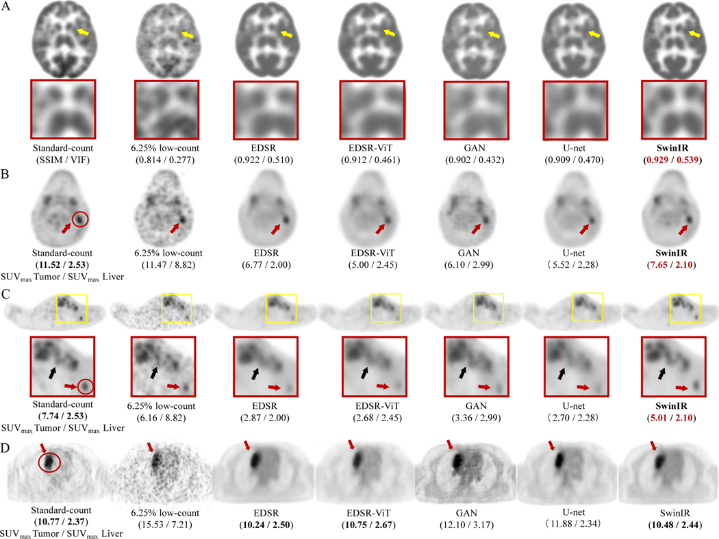
Figure 2: PET image comparison across five state-of-the-art AI algorithms on 6.25% low-count PET reconstruction.
(A) Representative 18F-FDG PET scan of a 29-year-old female patient with Hodgkin lymphoma (HL). The enlarged patches are shown on the second panel (yellow arrows: basal ganglia). The structural similarity index (SSIM) and visual information fidelity (VIF) metrics are presented under each PET image. (B) Representative 18F-FDG PET scan of a 14-year-old male patient with HL. The SUVmax of the lesion (delineated by red circle) and liver for this patient are shown under each PET image. (C) The same patient as (B). The small lesion (less than 1.5 cm3; 5mm < width <10mm; height > 10mm; red arrow ) is enhanced by SwinIR with the lesion-to-liver contrast of SUVmax retained. The lesions (black arrow) are also clearly depicted by SwinIR, in contrast with being blurred and mixed together by the other reconstructions. (D) Representative 18F-FDG PET scan of a 17-year-old female patient from the external Tübingen testing cohort. All AI algorithms successfully denoise the 6.25% low-count images and provide similar diagnostic conspicuity of the lesion (red circle; red arrows) as the standard-dose PET, demonstrating the model is generalizable across different institutions for all AI algorithms. SwinIR shows superiority in retaining lesion-to-liver contrast and structural fidelity.

