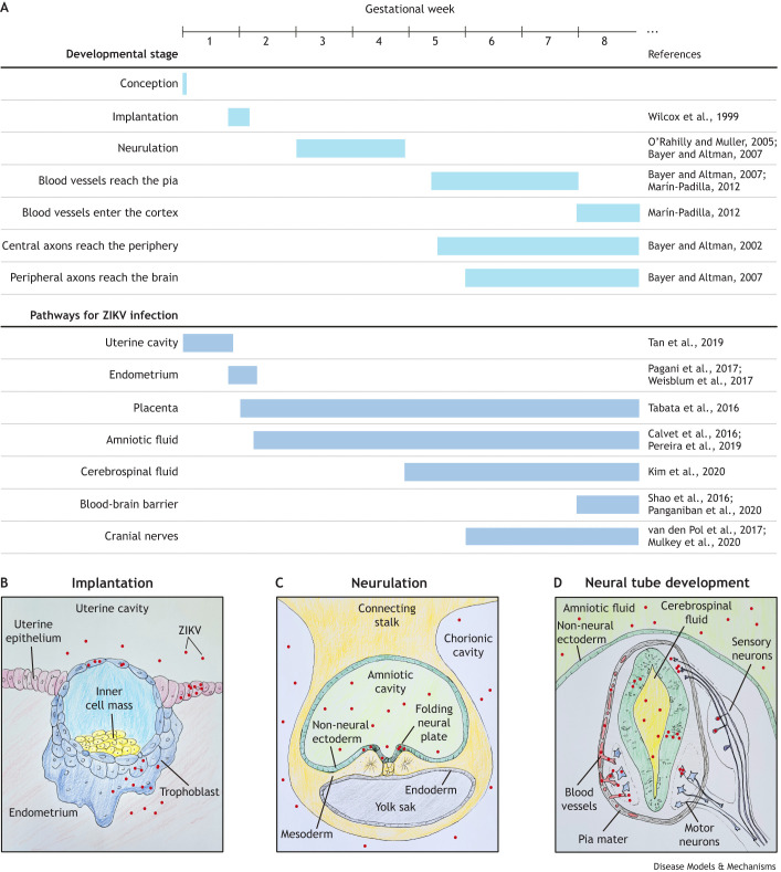
Fig. 1.
Human early embryogenesis milestones and the possible routes for Zika virus (ZIKV) infection and spread throughout the central nervous system (CNS). (A) Timeline of key human developmental milestones and the appearance of pathways for ZIKV transport to the embryonic CNS. Relevant references that support each milestone timing and infection pathway are provided in the right column. (B-D) Representative drawings of the development of routes for ZIKV transport in the early embryo. (B) Transverse section of the embryo implanting in the endometrium. After the blastocyst hatches from the zona pellucida, trophoblast cells come into direct contact with the uterine environment. ZIKV infects trophoblast cells and possibly reaches the embryonic cells in the inner cell mass. After the embryo implants in the endometrium, it loses its direct contact with the uterine cavity. However, ZIKV can infect endometrial cells. Therefore, ZIKV can infect trophoblast cells through the uterine cavity and through the endometrium. (C) Transverse section of the trilaminar embryonic disc surrounded by the amnion and yolk sac. Human neurulation starts at the third gestational week (GW). At this stage, neural cells are in contact with the amniotic fluid, which can also become infected. ZIKV in the amniotic fluid infects the neural plate cells but not the non-neural ectoderm. It does not display tropism for mesoderm or endoderm cells either. (D) Transverse section of the neural tube at the second month of gestation. After the non-neural ectoderm closes, the amniotic fluid is separated from the neural tube and is therefore not a possible direct infection pathway anymore. From this stage on, to infect the CNS, ZIKV has to cross through the barriers between the blood and the meninges (represented on the left side of the neural tube in red and gray, respectively), the blood and the cerebrospinal fluid, or the blood and the brain parenchyma (represented on the left side of the neural tube). Alternatively, ZIKV needs to be transported along the nerves that leave (motor neurons; blue) or enter (sensory neurons; purple) (both neuron types are represented on the right side of the neural tube) the CNS.

