Fig. 3.
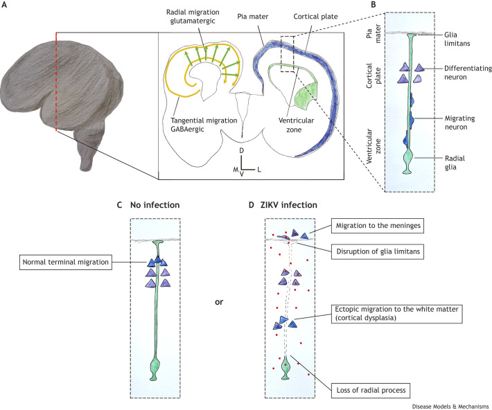
ZIKV-induced neuronal migration defects. (A) Schematic representation of a human brain at the 17th GW. The dashed red line indicates the position of the coronal section represented in the rectangle on the right. The schematic of the left hemisphere shows the two types of neuronal migration in the cortical plate: the tangential migration of inhibitory GABAergic neurons (yellow arrows) and the radial migration of excitatory glutamatergic neurons (green arrows). In the schematic of the right hemisphere, the proliferative layers are represented in green and the developing cortical plate is depicted in blue. The dashed line box indicates the localization of panels B-D. (B) At this stage of brain development (17th GW), radial glia form a physical support for the migration of newly generated neurons. The schematic shows that the neurons that previously migrated and are starting to differentiate reside closer to the basal process of the radial glia (where it attaches to the pia mater). (C) In non-infected developing brains, the migrating neurons pass through the previously formed cortical layers and detach from the radial glia closer to the pia, clustering in the cortical plate. (D) In ZIKV infection, the basal processes of radial glia can become deformed or are lost, diminishing cues for proper neuronal migration. Moreover, the loss of glia limitans (the radial glial foot in the pia) increases the ‘permeability’ of the zone between the brain parenchyma and the meninges, allowing developing neurons to ectopically migrate to the meninges. Additionally, loss of radial glia processes causes premature interruption of neuronal migration, resulting in ectopic accumulation in the white matter. D, dorsal; V, ventral; M, medial; L, lateral.

