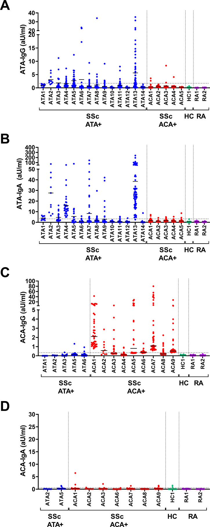
Figure 1.
IgG and IgA ATA and ACA in cell culture supernatants on in vitro PBMC stimulation. PBMC from patients with ATA+SSc, ACA+SSc, HC and RA were cultured on ex vivo isolation in 96-well flat bottom plates at a cell density of 2×10⁵/well in the presence of CD40L-expressing fibroblasts, IL-21 and BAFF. Culture supernatants were assessed for the presence of ATA-IgG (A), ATA-IgA (B), ACA-IgG (C) and ACA-IgA (D) by ELISA after 7 days of culture. Each column represents one patient, each dot one culture well. Lines depict median levels of secretion. Dotted line represents the cut-off based on the mean plus two times the SD of measurements of all controls. Average of 30 wells/donor (10–50 wells/donor). ACA, anti-centromere antibodies; ATA, anti-topoisomerase antibodies; BAFF, B cell activating factor; HC, healthy controls; IgG, immunoglobullin G; IL-21, interleukin 21; PBMC, peripheral blood mononuclear cells; RA, rheumatoid arthritis; SSc, systemic sclerosis.

