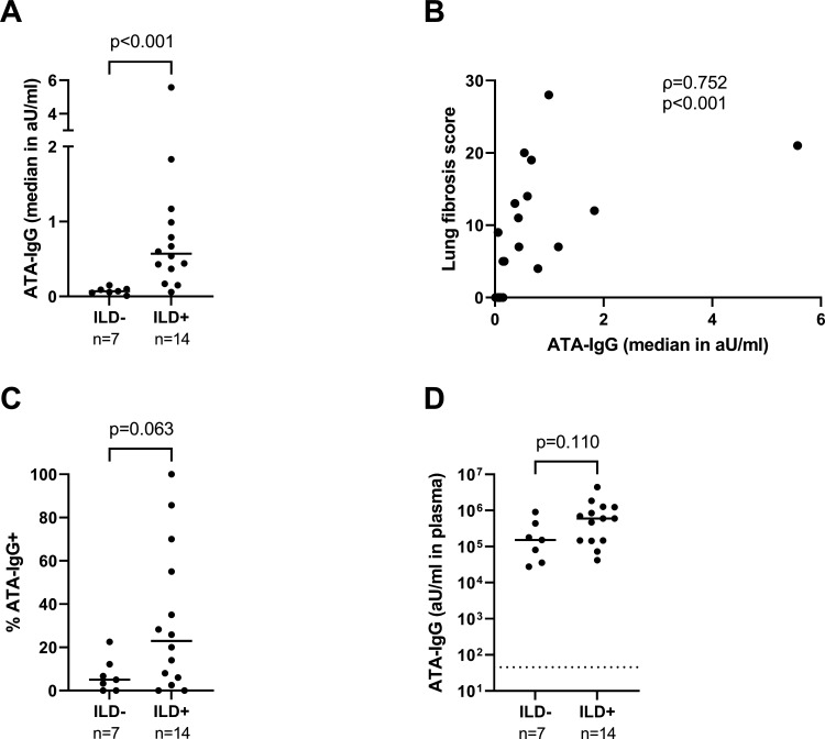
Figure 3.
Spontaneous secretion of ATA-IgG by PBMC in relation to the presence and severity of ILD. PBMC from ATA+SSc patients with ILD (n=14) or without ILD (n=7) were cultured without stimulation. ATA-IgG was measured in culture supernatants by ELISA after 7 days. (A) Levels of ATA-IgG (median in aU/mL) were compared between groups. (B) Levels of ATA-IgG produced spontaneously (median in aU/mL) in relation to the lung fibrosis score. (C) Percentage of wells positive for ATA-IgG were compared between groups. (D) ATA-IgG plasma levels in the same patients. Lines depict median values. Dotted line represents the cut-off based on the mean plus two times the SD of measurements of HC (n=11) and patients with RA (n=3). P values and Spearman correlation (ρ) are shown. ATA, anti-topoisomerase antibodies; HC, healthy controls; IgG, immunoglobulin G; ILD, interstitial lung disease; PBMC, peripheral blood mononuclear cells; SSc, systemic sclerosis.

