FIGURE 6.
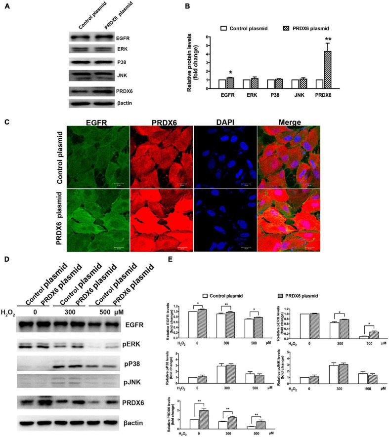
The effect of PRDX6 overexpression on EGFR and MAPKs in ARPE-19 cells. (A) Cells were infected with lentivirus containing the PRDX6 plasmid. Total EGFR, P38MAPK, ERK, JNK, and β-actin were detected by western blot. (B) Quantitative analysis of western blot results, *P < 0.05, **P < 0.01, compared to control. (C) Immunofluorescence staining of EGFR, PRDX6, and DAPI after ARPE-19 cells were infected with lentivirus containing a control plasmid or the pLV-Flag PRDX6 plasmid. Scale bar = 20 μm. (D) Cells were infected with lentivirus containing a control plasmid or the pLV-Flag PRDX6 plasmid for 48 h and were treated with 300 and 500 μM H2O2 for 6 h. EGFR, P38MAPK, ERK, JNK, and β-actin were detected by western blot. (E) Quantitative analysis of western blot results is shown in panel (D). All data were from three independent experiments, *P < 0.05, **P < 0.01.

