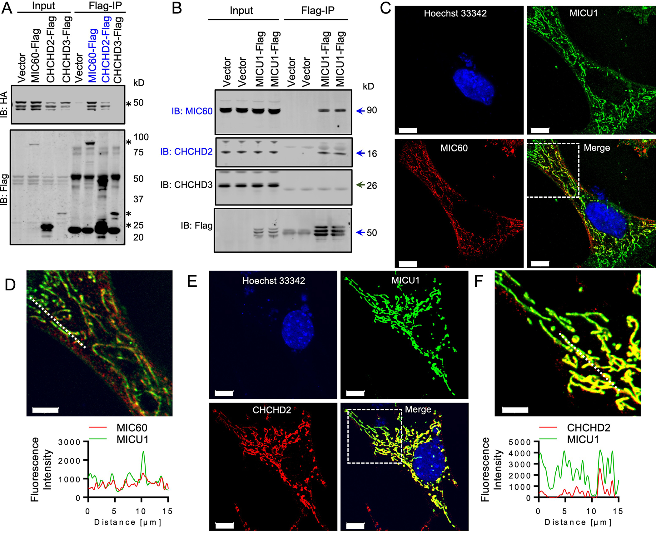
Fig. 3. MICU1 directly interacts with MICOS components.

(A) MICU1-HA and FLAG-tagged MICOS components were co-expressed in MICU1−/− HEK293T cells. FLAG-immunoprecipitates (IPs) were probed with FLAG and HA antibodies to detect the interaction between MICU1 and MICOS components. Asterisk (*) indicate the bands for the specific Flag-tagged MICOS components or MICU1-HA. The blue font indicates positive interactions. Western blots are representative of 3 independent experiments. (B) FLAG immunoprecipitates from MICU1−/− HEK293T cells reconstituted with MICU1-FLAG were immunoblotted for endogenous MICOS components. Arrows indicate the specific protein bands. The blue font indicates positive interactions. Western blots are representative of 3 independent experiments. (C-F) MICU1-FLAG expressing MEFs were imaged for FLAG and MIC60 (C, D) or FLAG and CHCHD2 (E, F) and line scans of MICU1, MIC60 and CHCH2 were performed. Images are representative of 3 independent experiments. Scale bar = 10μm in (C) and (E) and 5μm in (D) and (F).
