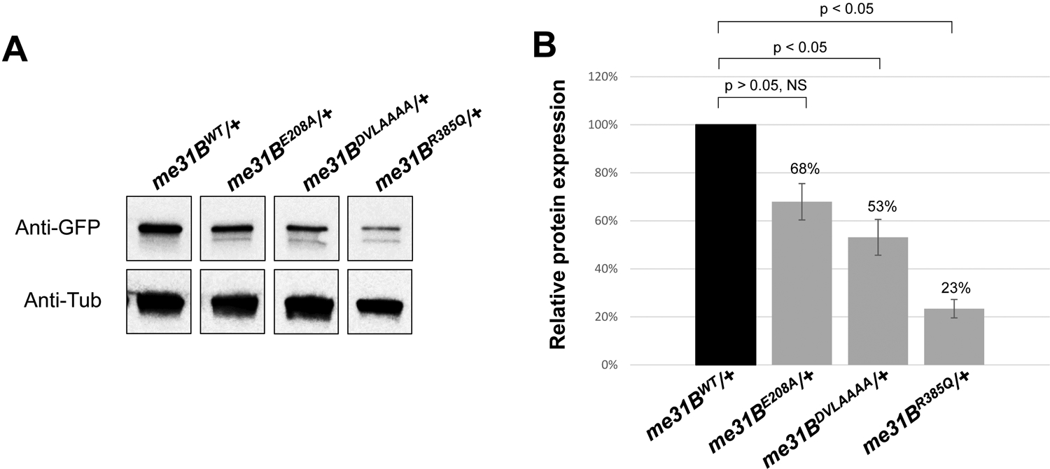
Figure 3. Western blot quantification of mutant Me31B protein levels in me31BE208A, me31BDVLAAAA, and me31BR385Q heterozygous strains.

(A) Anti-GFP Western blots were used to quantify the Me31BWT-GFP, Me31BE208A-GFP, Me31BDVLAAAA-GFP, and Me31BR385Q-GFP proteins in the ovaries of the corresponding heterozygous strains. Anti-tubulin Western blots were used as loading controls. The expression levels of Me31BE208A-GFP, Me31BDVLAAAA-GFP, and Me31BR385Q-GFP proteins were lower than the Me31BWT control. The images shown are representative images of three biological replicates. The additional, uncropped biological replicate images are presented in Supplementary Figure 1. (B) The Me31BE208A-GFP, Me31BDVLAAAA-GFP, and Me31BR385Q-GFP protein levels are 68% (p > 0.05), 53% (p < 0.05), and 23% (p < 0.05) relative to the Me31BWT-GFP control, respectively. Western blot image analysis was performed with ImageJ and protein quantification was normalized by using the alpha-tubulin proteins. NS, not statistically significant. Error bar represents the standard error of the mean.
