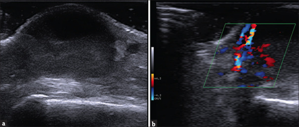
Figure 2.
Ultrasound of the nodular formation of the dorsal hand using a high-frequency linear probe [SL3235 appleprobe – Esaote] (a) Deep to the epidermal line, starting from the upper part of the papillar dermis, there we can see the well limited the cutaneous abscess. On the upper part of the lesion a clear anechoic area corresponding to the liquid-exudative part; down it a less well-demarcated area with a non-homogeneous and hypoechoic aspect, with internal echoes, corresponding to colliquative material and debris. On the borders are visible residual collagen bundles of reticular dermis. (b) Colordoppler imaging of the lesion (40% Gain). We note the heterogeneous and bidirectional speed flow with different spot origins of the signal, indicative of strong vascular activity caused by an inflammatory component

