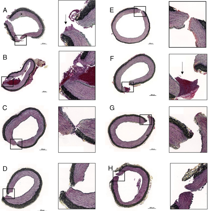
Figure 4.

Histopathologic samples of each gauze demonstrating some degree of myocyte degeneration (black asterisk) and fibrin deposition (black arrows). QuikClot Combat Gauze (A), BL-IX (B), TAC (C), ChitoSAM 100 (D), CR (E), EVARREST Fibrin Sealant Patch (F), ChitoGauze XR Pro (G), XS (H). Scale bars on low magnification images are 0.5 mm.
