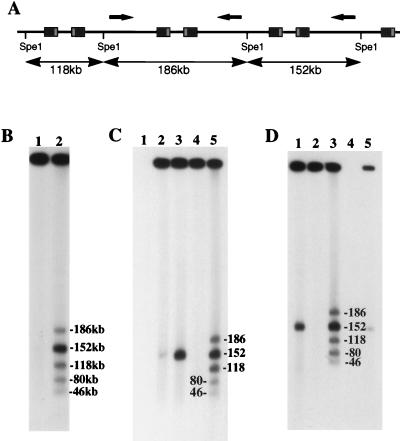
FIG. 1.
Analysis of fragments generated by SpeI digestion of high-molecular-weight HSV DNA purified from infected cells by FIGE. (A) Schematic representation of SpeI fragments generated when adjacent L segments are in opposite orientations (arrows). (B) Southern blot of SpeI fragments from SC16-infected cells. Bands were sized, as marked, by comparison with concatemeric λ phage DNA and correspond to expected fragments (lane 2). Terminal fragments (80 and 46 kb) corresponded exclusively to L-segment termini, as previously described (34). Undigested material (lane 1) contained no residual genomic-length HSV DNA. (C) Southern blot showing fragments indicative of L-segment inversion (lane 5) in high-molecular-weight DNA purified from ts+7-infected cells. Terminal fragments were from the L segment only (80 and 46 kb). Removal of unit-length HSV DNA (152 kb) was shown to be complete by comparing undigested samples before and after FIGE purification of high-molecular-weight material (lanes 3 and 4, respectively). The migration of unit-length genomes was marked by using total DNA extracted 5.5 h after infection of cells with SC16 (lane 2). There was no background hybridization to uninfected-cell DNA (lane 1). (D) Southern blot showing L-segment inversion and L-segment termini in high-molecular-weight DNA from R7713-infected cells. Comparison between undigested total infected-cell DNA (lane 1) and high-molecular-weight DNA (lane 2) showed that unit-length genomes were removed from the analyzed sample. Lane 4, not loaded; lane 5, total undigested DNA 4 h after SC16 infection.

