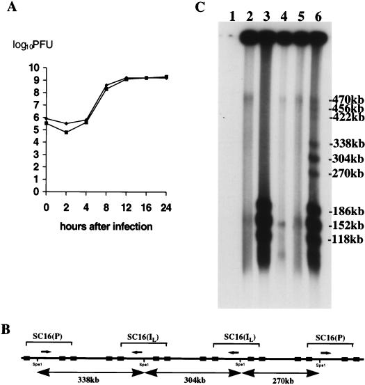
FIG. 2.
(A) Single-step growth curves (10 PFU/cell) of SC16 and 5B8 in Vero cells. (B) SC16-5B8 recombination; expected SpeI fragments from concatemers comprising L segments of 5B8 flanked by SC16. (C) FIGE and Southern blot analysis of fragments generated by SpeI digestion of high-molecular-weight infected-cell DNA (17 h after infection). Lane 1, uninfected Vero cells; lanes 2 and 3, high-molecular-weight SC16 DNA; lane 4, high-molecular-weight 5B8; lanes 5 and 6, mixed infection with SC16 and 5B8. Samples in lanes 3, 4, and 6 were treated with SpeI. Fragments, as marked, were sized by comparison with high-molecular-weight markers. A small amount of residual genomic-length DNA was detected in the sample loaded into lane 4. The filter was autoradiographed for 1.5 h.

