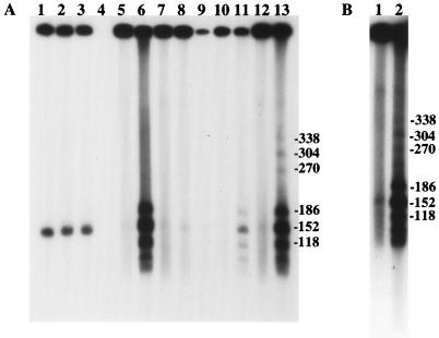
FIG. 3.
(A) Analysis of total and high-molecular-weight DNAs recovered from infected cells at various times after infection. Lanes 1 to 3, total infected-cell DNA recovered from cells infected with SC16, 5B8, or both viruses, respectively, demonstrating the appearance of unit-length progeny DNA (152 kb), 6 h after infection. Lane 4, uninfected-cell DNA; lanes 5 and 6, SC16; lanes 7 and 8, 5B8; lanes 9 to 13, mixed infection. Agarose plugs loaded into lanes 6, 8, 9, 11, and 13 were treated with SpeI. Lanes 5 to 8 and 12 and 13 show results 13.5 h after infection. Lanes 10 and 11 show results 6 h after infection. Lane 9, 4 h after infection, contained insufficient DNA for detection of putative recombinants. The filter was autoradiographed for 15 min. (B) Reanalysis of samples made 6 h after infection with SC16 and 5B8. Lane 1, uncut; lane 2, SpeI cut. The filter was autoradiographed for 5 h.

