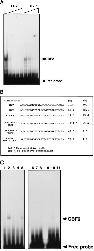
FIG. 3.
Affinities of HVP and RhEBV EBNA2 enhancers for CBF2. Nuclear extracts from CA46 cells were mixed with 0-, 2.5-, 5-, 25-, and 125-fold molar excesses of the different unlabeled mutant oligonucleotides. A fixed amount of 32P-EBV CBF2 binding oligonucleotide was then added to the shift reaction mixtures. The reaction mixtures were separated on 4.5% nondenaturing polyacrylamide gels, dried, and autoradiographed. Amounts of CBF2 complex were quantified with a PhophorImager (Molecular Dynamics). (A) The EMSA gel shows binding of nuclear extract containing CBF2 to an EBV 36-mer oligonucleotide probe from positions −339 to −368 alone or with increasing (triangle) amounts of the indicated equivalent EBV or HVP cold competitor oligonucleotide. (B) Summary of competition results. The sequences shown represent the central 30 bp of the 36-mer oligonucleotide. The crucial sequences for CBF2 binding are in bold, with the mutational changes underlined. The numbers in column a are concentrations of unlabeled oligonucleotide required for 50% competition. The numbers in column b are percentages representing the ability of each oligonucleotide to compete for CBF2 relative to that of the wild-type element, which was set at 100%. (C) Direct binding of HVP CBF2 wild-type and mutant oligonucleotides. Nuclear extracts from CA46 cells were incubated with radiolabeled HVP wild-type and mut.1 and mut.2 CBF2 oligonucleotides in the presence or absence of a 100 M excess of competitor oligonucleotide and analyzed by EMSA. For lanes 1 to 5, the HVP CBF2 probe was used; for lanes 6 to 8, the HVP mut.1 CBF2 probe was used; and for lanes 9 to 11, the HVP mut.2 probe was used. Lanes 1, 6, and 9, probe only; lanes 2, 7, and 10, CA46 extract; lane 3, CA46 extract and cold HVP CBF2 oligonucleotide; lanes 5 and 8, CA46 and cold HVP mut.1 CBF2 oligonucleotide; lanes 4 and 11, CA46 and cold HVP mut.2 CBF2 oligonucleotide. Results are averages of values from three independent experiments.

