
Figure 3.
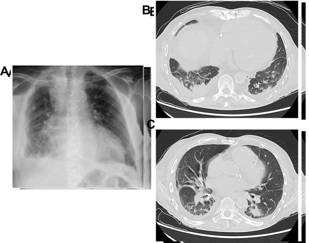
Elderly patient with a high clinical suspicion of pneumonia. Because of overlapping pulmonary oedema and effusion, X-ray (A) was non-conclusive. In a subsequently performed CT scan, however, small consolidations in (B, C) right lower lobe are visible.