. 2020 Sep 15;133(18):jcs247593. doi: 10.1242/jcs.247593

Fig. 4.

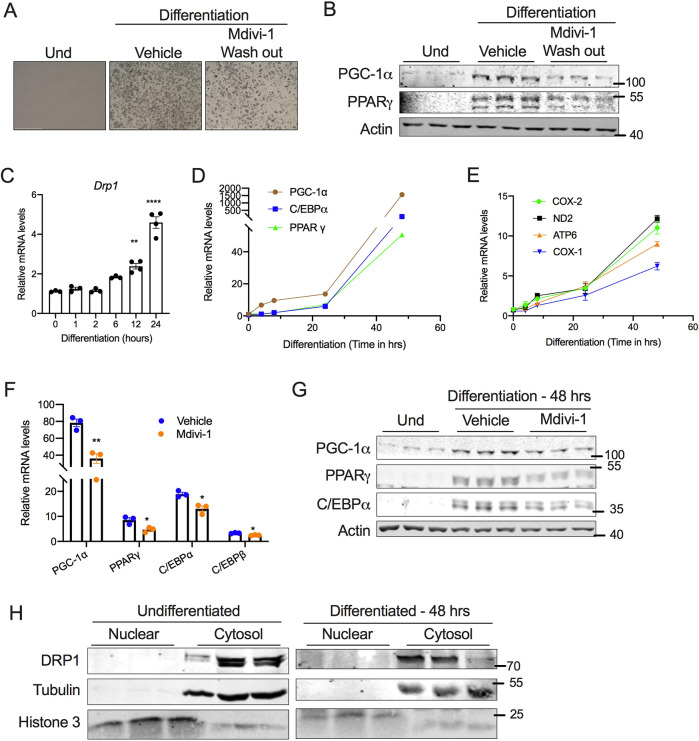
Fig. 4. DRP1 is indispensable for the induction of beige adipocyte differentiation. (A) Representative images for SVF cells differentiated into beige adipocytes in the absence or presence of 10 µM mdivi-1 during the induction period (48 h), followed by wash out and continued differentiation on maintenance medium without mdivi-1 for the next 6 days. Scale bars: 500 µm. (B) Immunoblots for expression of adipogenesis-related proteins in cells treated as described in A. (C–E) qPCR analysis assessing the time-dependent change in expression of (C) Drp1, (D) adipogenesis-related genes and (E) mitochondrial markers during the early phase of beige adipocyte differentiation. (F) qPCR and (G) western blot analysis of genes involved in adipogenesis assessed at 48 h following the induction of SVF cell differentiation in the presence or absence of 10 µM mdivi-1. (H) Immunoblots of DRP1 in nuclear and cytosolic fractions of SVF cells assessed at 48 h following induction of differentiation. In C–F, β-actin was used to normalize gene expression. In B and G, actin is shown as a loading control. In H, tubulin and histone 3 are shown as cytosolic and nuclear loading controls, respectively. Quantitative data are presented as mean±s.e.m. (*P<0.05, **P<0.01, ****P<0.0001). Und, undifferentiated.

DRP1 is indispensable for the induction of beige adipocyte differentiation. (A) Representative images for SVF cells differentiated into beige adipocytes in the absence or presence of 10 µM mdivi-1 during the induction period (48 h), followed by wash out and continued differentiation on maintenance medium without mdivi-1 for the next 6 days. Scale bars: 500 µm. (B) Immunoblots for expression of adipogenesis-related proteins in cells treated as described in A. (C–E) qPCR analysis assessing the time-dependent change in expression of (C) Drp1, (D) adipogenesis-related genes and (E) mitochondrial markers during the early phase of beige adipocyte differentiation. (F) qPCR and (G) western blot analysis of genes involved in adipogenesis assessed at 48 h following the induction of SVF cell differentiation in the presence or absence of 10 µM mdivi-1. (H) Immunoblots of DRP1 in nuclear and cytosolic fractions of SVF cells assessed at 48 h following induction of differentiation. In C–F, β-actin was used to normalize gene expression. In B and G, actin is shown as a loading control. In H, tubulin and histone 3 are shown as cytosolic and nuclear loading controls, respectively. Quantitative data are presented as mean±s.e.m. (*P<0.05, **P<0.01, ****P<0.0001). Und, undifferentiated.