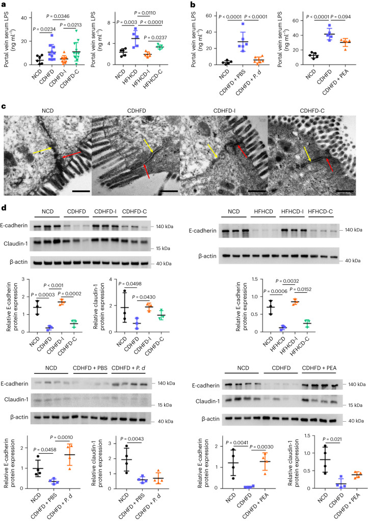
Fig. 5. Inulin, P. distasonis and pentadecanoic acid restored gut barrier function.
a, Portal vein serum LPS in CDHFD- or HFHCD-induced NASH mouse models treated with inulin or cellulose. Between 7 and 12 mice were used in each group in the first model: NCD (n = 7), CDHFD (n = 10), CDHFD-I (n = 10) and CDHFD-C (n = 12). Five mice were used in each group in the second model. b, Portal vein serum LPS in a CDHFD-induced NASH model treated with P. distasonis or pentadecanoic acid. Between five and seven mice were used in each group. c, Representative TEM images of the gut epithelium from mice fed NCD, CDHFD, CDHFD-I or CDHFD-C. Scale bars, 400 nm. Red and yellow arrows indicate a tight junction and adherence junction, respectively. d, Tight and adherence junction marker expression in NASH mouse models treated with inulin, cellulose, P. distasonis (P. d) or PEA, n = 3–4 per group. a,b,d, Data are presented as biological replicates ± s.d. P value obtained by one-way ANOVA with Fisher’s LSD.

