Fig. 1.
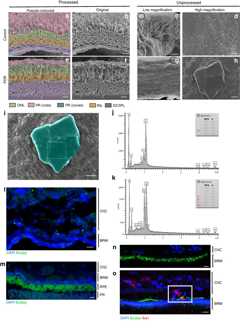
Increased retinal cholesterol levels and cholesterol crystallisation in vivo result in elevated inflammation, cell death and vascular permeability. (a–h) Pseudo-coloured and non-coloured processed and unprocessed tissue samples from a non-diabetic control donor and a diabetic donor with PDR. A representative CC is shown in (h) for the diabetic donor with PDR. Black circles in (c) and (g) indicate the general vicinity of the areas where the high-magnification images were taken. (i–k) EDS composition analysis of a representative crystal (pseudo-coloured in teal) and surrounding tissue from the PDR retinal section. (l, m) Tissue samples from a non-diabetic control donor (l) and a diabetic donor without diabetic retinopathy (m) stained with Bodipy (green) and DAPI (blue). (n, o) Proliferative diabetic choroid from a diabetic donor with PDR stained with Bodipy (green), DAPI (blue) and Iba1 (red). Scale bars: 50 µm (a, b, e, f), 20 µm (c, g), 10 µm (d, h, i), 50 µm (l, m, n), 15 µm (o). BRM, Burch’s membrane; ChC, choroidal capillaries; GC/IPL, ganglion cell inner plexiform layer; INL, inner nuclear layer; ONL, outer nuclear layer; PR, photoreceptors; RPE, retinal pigment epithelium

