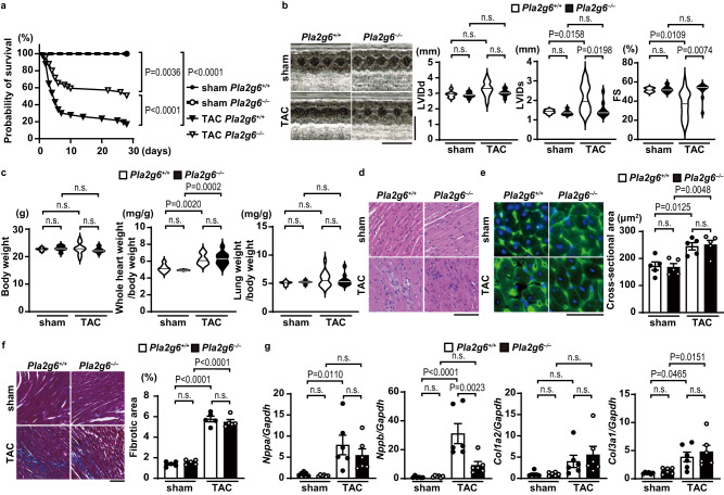
Fig. 1. Cardiac phenotypes of cardiac-specific iPLA2β-deficient mice after pressure overload.
The Pla2g6+/+ and Pla2g6–/– mice were subjected to transverse aortic constriction (TAC) and then analyzed 5 days after the operation. a Survival ratio after TAC. n = 17 (sham-operated Pla2g6+/+), 13 (sham-operated Pla2g6–/–), 51 (TAC-operated Pla2g6+/+), and 51 (TAC-operated Pla2g6–/–). The log-rank test was used for survival analysis. Sham Pla2g6+/+ versus TAC Pla2g6+/+ P = 0.0000001, TAC Pla2g6+/+ versus TAC Pla2g6–/– P = 0.0002. b Representative images of transthoracic M-mode echocardiographic tracing (scale bars, 0.2 s and 5 mm, respectively) and the echocardiographic parameters of the mice. n = 7 (sham-operated Pla2g6+/+), 7 (sham-operated Pla2g6–/–), 14 (TAC-operated Pla2g6+/+) and 10 (TAC-operated Pla2g6–/–). LVIDd and LVIDs, end-diastolic and end-systolic left ventricular (LV) internal dimensions; FS, fractional shortening of LV. c Physiological parameters of the mice. n = 7 (sham-operated Pla2g6+/+), 7 (sham-operated Pla2g6–/–), 14 (TAC-operated Pla2g6+/+), and 10 (TAC-operated Pla2g6–/–), biologically independent samples. d Representative images of the hematoxylin—eosin-stained heart sections. Experiment was repeated five times independently with similar results. Scale bar, 100 μm. e Representative images of the wheat germ agglutinin (green)-stained heart sections. Scale bar, 50 μm. Blue, DAPI. The graph shows the cross-sectional area of cardiomyocytes (n = 5, biologically independent samples). f Representative images of Azan—Mallory-stained heart sections. Scale bar, 100 μm. The graph shows the ratio of the fibrotic area to whole heart section (n = 5, biologically independent samples. Sham Pla2g6+/+ versus TAC Pla2g6+/+ P = 0.0000000002, sham Pla2g6–/– versus TAC Pla2g6–/– P = 0.000000001). g Expression levels of mRNAs related to cardiac remodeling. (n = 6, biologically independent samples. Sham Pla2g6+/+ versus TAC Pla2g6+/+ in Nppb/Gapdh P = 0.00006). Data were normalized to the Gapdh content and are expressed as the fold increase over the levels in the sham-operated Pla2g6+/+ group. Open and closed bars indicate Pla2g6+/+ and Pla2g6−/−, respectively. In violin plots, solid lines show median. In bar graphs, data are expressed as the mean ± SEM. The data were evaluated by one-way ANOVA, followed by Tukey-Kramer’s post-hoc test. Source data are provided as a Source Data file.

