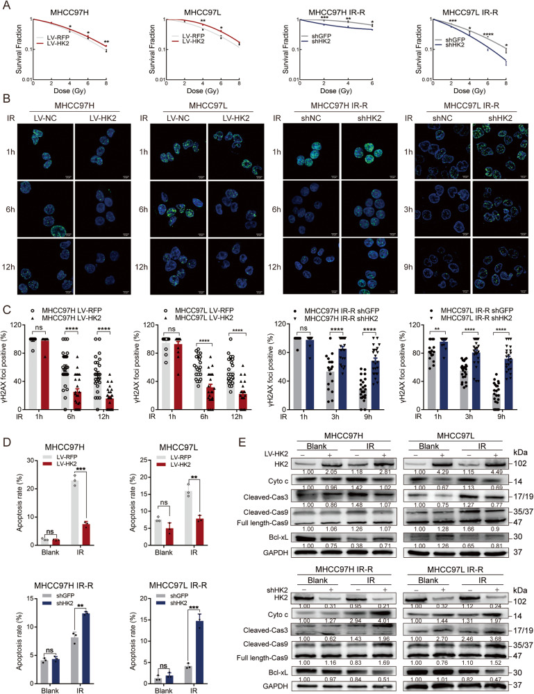
Fig. 2. HK2’s role in proliferation and IR-induced apoptosis.
A Colony formation assays of cells with different HK2 expressions after exposure to indicated doses of radiation. Error bars are the SEM of at least three independent replicates. B, C Cells were treated with 8 Gy radiation and then stained and quantified at the indicated times with antibodies to pH2AX-Ser139. Error bars are the SEM of at least ten independent replicates. Scale bar 20 μm. D Statistics of apoptosis in indicated cell lines with different HK2 status with or without 8 Gy radiation, respectively (n = 3). *p < 0.05, **p < 0.01, ***p < 0.001, ****p < 0.0001, based on Students’ t-test. E Western blotting of HK2, Cyto c, Cleaved Caspase 3, Caspase/Cleaved Caspase 9, and Bcl-xL in indicated groups.

