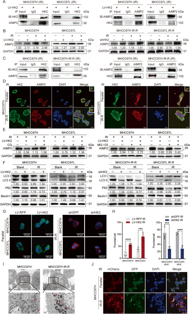
Fig. 4. HK2 co-localizes with AIMP2 post IR.
A Co-immunoprecipitation (Co-IP) identified the interaction between HK2 and AIMP2 in MHCC97H LV-HK2 and MHCC97L LV-HK2 post 8 Gy radiation. B Protein levels of AIMP2 in HCC cell lines with different HK2 expressions by facilitating western blotting post 8 Gy radiation. C Co-IP identified the interaction between HK2 and AIMP2 in MHCC97H and MHCC97H IR-R post 8 Gy radiation. D IF analysis of the co-localization of HK2 (green) and AIMP2 (red) in MHCC97H, MHCC97L, MHCC97H IR-R, and MHCC97L IR-R post 8 Gy radiation. Scale bar 20 μm. E MG-132 (20 μM, 24 h) and CQ (5 μM, 24 h) individually treatment in MHCC97H and MHCC97L cells with or without HK2 overexpression post 8 Gy radiation. F Western blotting of autophagy-related protein including Beclin1, LC3 I/II, P62 in indicated groups with different HK2 and AIMP2 status post 8 Gy radiation. G, H IF analysis and quantification of the LC3 II (green) in MHCC97H and MHCC97L cells with or without HK2 overexpression, and LC3 II (red) in MHCC97H IR-R, MHCC97L IR-R cells with or without HK2 knockdown post 8 Gy radiation after 48 h (n = 5). Scale bar 40 μm. I TEM of indicating cells post 8 Gy radiation after 48 h. J IF analysis of autophagosomes in indicated post-IR cells which transfected with a plasmid expressing mCherry-GFP-LC3 II. Scale bar 20 μm. Data were represented as mean ± SEM. *p < 0.05, **p < 0.01, ***p < 0.001, ****p < 0.0001.

