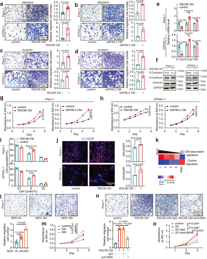
Fig. 4. Schwann cells promote malignant progression in PDAC via MDK.
a–d Effects of RSC96 CM (a, c) and sNF96.2 CM (b, d) on migration (a, b) and invasion (c, d) of Panc-1 and CFPAC-1 cells were assessed by Transwell and Matrigel invasion assays. n = 10, 15, 16 (a) or 5 (b–d) representative pictures over three independent experiments. Scale bar, 200 µm. e, f The relative expression levels of EMT markers in Panc-1 and CFPAC-1 cells cultured with RSC96 CM were detected by qPCR (e) and western blotting (f). Data were representative of n = 3 independent experiments. g–j Effects of RSC96 CM and sNF96.2 CM on the cell proliferation of Panc-1 and CFPAC-1 cells were assessed by CCK-8 (g, h), flow cytometry (i), and EdU assays (j). Scale bar, 100 µm. k QuSAGE scores of control and SC-CM associated signature in perineural tier 1‒4 and other regions in the neuro-stroma niche. The signatures were based on the bulk RNA-seq of CFPAC-1. l, m Effects of recombinant human MDK on migration (l) and proliferation (m) of Panc-1 cells were assessed by Transwell (l) and CCK-8 assay (m). Scale bar, 200 µm. n, o. Effects of MDK neutralization antibodies on migration (n) and proliferation (o) of Panc-1 cells cultured with RSC96 CM were assessed by Transwell (n) and CCK-8 (o) assays. Scale bar, 200 µm. e, g–j, m, n Data were the mean ± s.d. of n = 3 independent experiments. l, o Data were the mean ± s.d. of n = 5 independent experiments. Statistical analysis: unpaired two-sided t-test (a–e, i, j, l, n); two-way ANOVA (g, h, m, o). Source data are provided as a Source Data file.

