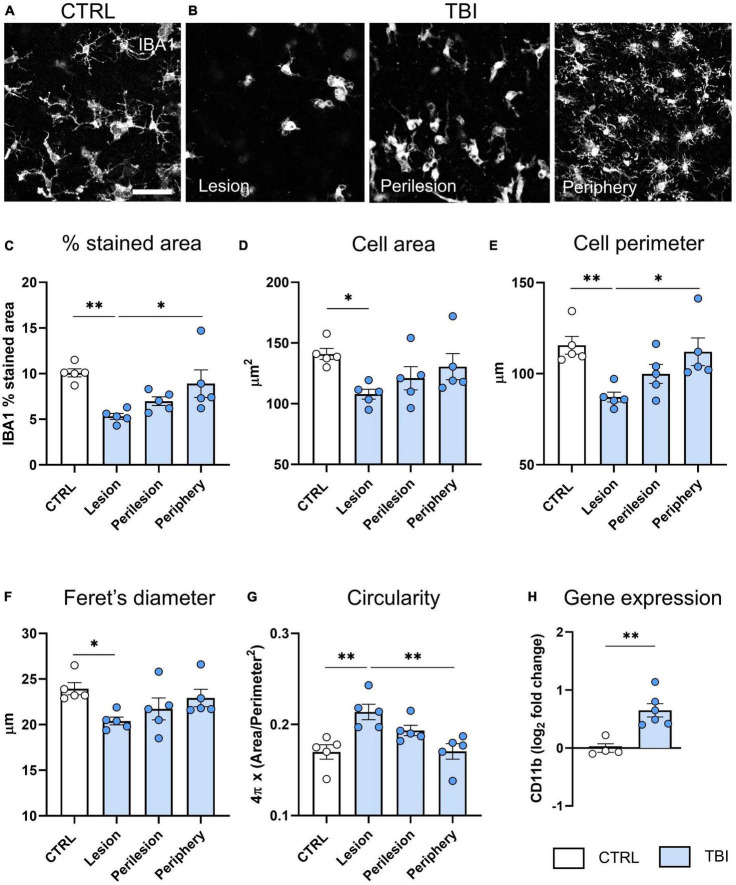
FIGURE 4.
Evaluation of TBI induced microglia changes 48 h after injury. (A) Representative confocal images of IBA-1 staining in CTRL slices and (B) in the lesion, perilesion, and periphery of TBI slices. Since Iba1 staining was uniform in CTRL slices throughout concentric areas, results are shown as the average of the 12 ROIs. (C) Quantification of Iba-1% stained area, and morphometric cell parameters: (D) mean cell area, (E) perimeter, (F) Feret’s diameter and (G) circularity. (H) Gene expression analysis of microglial CD11b marker in cortical slices. Data are mean ± SEM. (C–G) One-way ANOVA, followed by Tukey’s multiple comparisons test, (H) t-test. *p < 0.05 **p < 0.01. Bar = 50 μm.

