
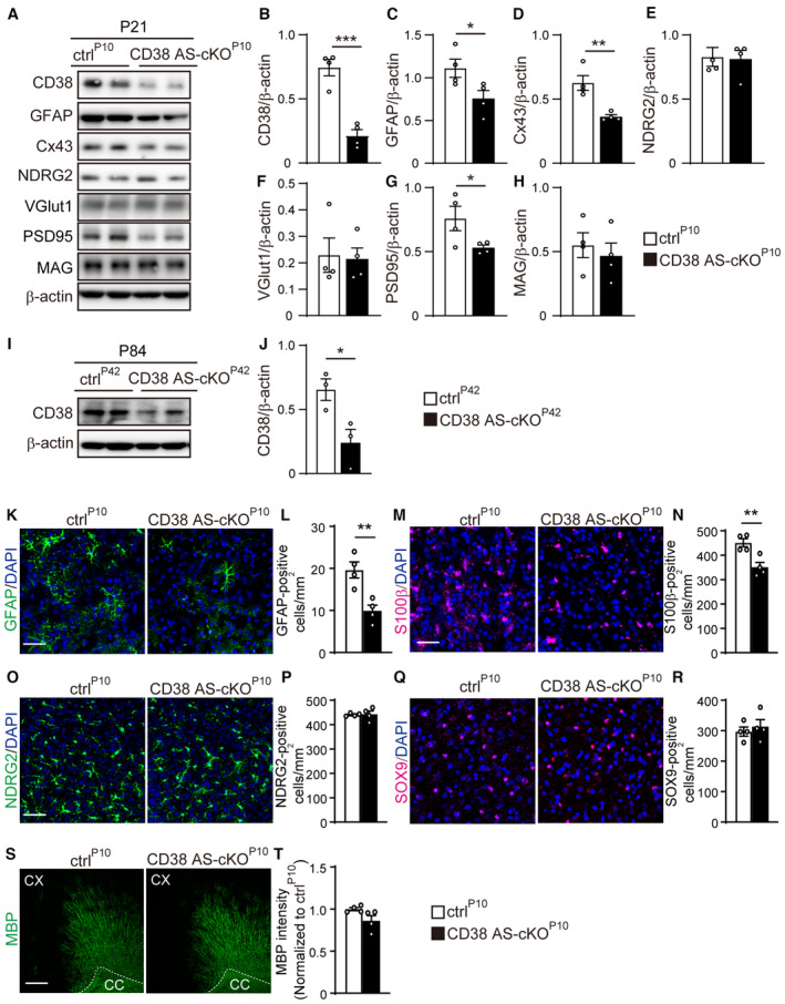
Figure EV2. Astrocyte‐specific deletion of CD38 decreases expression of GFAP, s100β and Cx43 in the developing mPFC.
-
ARepresentative western blots of CD38, GFAP, Cx43, NDRG2, VGlut1, PSD95, MAG, and β‐actin in the mPFC of ctrlP10 and CD38 AS‐cKOP10 mice at P21 (two animals per genotype are shown).
-
B–HRelative optical densities of CD38, GFAP, Cx43, NDRG2, VGlut1, PSD95, MAG, and β‐actin normalized to the loading control β‐actin (n = 4 animals per genotype, two‐tailed unpaired Student's t‐test).
-
IRepresentative western blots of CD38 and β‐actin in the mPFC of ctrlP42 and CD38 AS‐cKOP42 mice at P84 (two animals per genotype are shown).
-
JBar graphs depict the relative optical density of CD38 and β‐actin normalized to the loading control β‐actin (n = 3 animals per genotype, two‐tailed unpaired Student's t‐test).
-
K–T(K, M, O, Q, S) Immunohistochemistry for GFAP, s100β, NDRG2 and SOX9 in the mPFC and MBP in the motor cortex of ctrlP10 and CD38 AS‐cKOP10 mice at P21. Nuclei were counterstained with DAPI. Scale bars, 50 μm (K, M, O, and Q), and 100 μm (S). (L, N, P, R, T) Quantification of astrocyte‐specific protein‐positive cells and MBP intensity (n = 4 animals per genotype, two‐tailed unpaired Student's t‐test).
Data information: Data represent means ± SEM. *P < 0.05, **P < 0.01, ***P < 0.001.
Source data are available online for this figure.