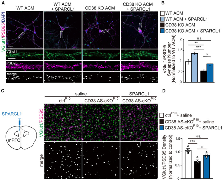
Figure 5. Astroglial CD38 promotes synapse formation through SPARCL1.

- Representative images of cortical neurons cultured for 14 DIV in WT ACM, WT ACM with SPARCL1, CD38 KO ACM, and CD38 KO ACM with SPARCL1 proteins. The insets show individual channels for VGlut1 (green) and PSD95 (magenta) staining, as well as the merged image. Nuclei were counterstained with DAPI. Scale bars: 20 μm (main image) and 10 μm (inset).
- Quantification of synapse numbers in cortical neurons (n = 40 to 55 cells per condition from five independent cultures, one‐way ANOVA followed by Tukey–Kramer test).
- SPARCL1 protein was stereotactically injected directly into layer II/III of the CD38 AS‐cKOP10 mPFC at P15. Immunohistochemistry for VGlut1 (green) and PSD95 (magenta) in the mPFC of ctrlP10 and CD38 AS‐cKOP10 mice at P18. The lower panels show the colocalized VGlut1 and PSD95 puncta. Scale bar, 10 μm.
- Quantification of synapse numbers in CD38 AS‐cKOP10 mPFC (n = 4 animals per condition, Kruskal–Wallis test followed by Dunn's test).
Data information: Data represent means ± SEM. N.S., no significance; *P < 0.05, **P < 0.01, ***P < 0.001.
