Figure 2.

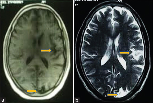
(a) T1W image showing hypointense lesions. (b) Corresponding T2W images show hyperintense lesions involving the left frontal cortical gray matter, left posterior occipital lobe, and adjoining sulcal spaces (indicated by yellow arrows)

(a) T1W image showing hypointense lesions. (b) Corresponding T2W images show hyperintense lesions involving the left frontal cortical gray matter, left posterior occipital lobe, and adjoining sulcal spaces (indicated by yellow arrows)