Figure 3.

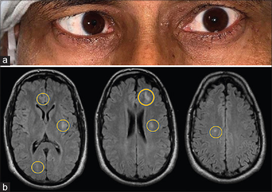
(a) Patient 2 with right eye esotropia. (b) T2/FLAIR images showing multiple hyperintense lesions involving the bilateral frontoparietal lobe, and left temporal lobe (indicated by yellow circles)

(a) Patient 2 with right eye esotropia. (b) T2/FLAIR images showing multiple hyperintense lesions involving the bilateral frontoparietal lobe, and left temporal lobe (indicated by yellow circles)