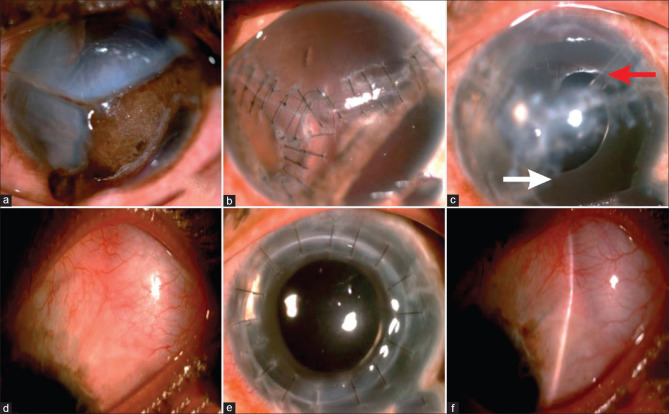
Figure 1.
(a-f) Pre- and Post-operative images (a): Left eye rupture with triradiate tear of cornea extending to limbus, uveal tissue prolapse. (b) Sealed globe with 10-0 nylon interrupted sutures, reduced corneal edema, iris remnants, and aphakia at 2 months post-primary repair. (c) Aniridia SFIOL with Ahmed glaucoma valve (AGV tube marked with red arrow, aniridia IOL with white arrow). (d) Diffuse bleb seen supero-temporally. (e) Post-penetrating keratoplasty, with a clear graft. (f) Well-functioning diffused bleb with controlled intraocular pressure

