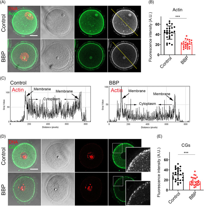
FIGURE 6.

Benzyl butyl phthalate (BBP) exposure affects the dynamics of Actin and cortical granules. (A) Germinal vesicle (GV) oocytes from each group were stained by phalloidin, and representative images of actin signal in control and BBP‐exposed oocytes. Green, Actin; red, DNA. (B) The average fluorescence intensity of actin was significantly decreased in BBP‐exposed oocytes comparing with that in the control oocytes. Results are expressed as mean ± SD; experiments were repeated at least three times (NS, not significant; *p < 0.05; **p < 0.01; ***p < 0.001). (C) Both cytoplasmic actin and cortical actin showed lower fluorescence intensity after BBP exposure. (D) GV oocytes from each group were stained by lens culinary agglutinin‐FITC. The representative images of CGs signal in control and BBP‐exposed oocytes. Green, CGs; red, DNA. (E) The fluorescence intensity of cortically distributed CGs was significantly decreased in BBP‐exposed oocytes comparing with that in the control oocytes. Results are expressed as mean ± SD; experiments were repeated at least three times (NS, not significant; *p < 0.05; **p < 0.01; ***p < 0.001).
