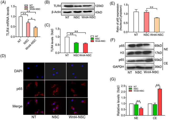
FIGURE 5.

Wnt4‐modified NSCs promote M2 polarization through suppressing of TLR4/NF‐κB signal pathway. (A) RT‐qPCR analysis of expressions of TLR4 in macrophages co‐cultured with NSCs, n = 3. (B) Western Blot analysis of expression of TLR4 in macrophages co‐cultured with NSCs, n = 3. (C) Quantification of western blot data in panel B, n = 3. (D) Immunofluorescence analysis of expression of p65 in macrophages co‐cultured with NSCs, n = 3, bar: 10 μm. (E) Quantification of immunofluorescence data in panel D, n = 3. (F) Western blot analysis of expression of p65 in nuclear extract (NE) and cytosolic extract (CE) of macrophages co‐cultured with NSCs. (G) Quantification of western blot data in panel E, n = 3. (The data are presented as the means ± SD from one representative experiment of three independent experiments performed in triplicate. **p < 0.01 compared between groups; *p < 0.05 compared between groups.)
