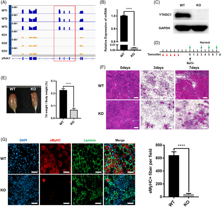
FIGURE 1.

Mice with satellite cell‐specific deletion of Ythdc1 are unable to regenerate skeletal muscles normally. (A) Expression of Ythdc1 exons in SCs from wild type (WT) and knockout (KO) mice based on RNA‐seq data (WT, n = 3; KO, n = 3). (B) Quantitative real‐time PCR was performed to detect deleted exons of Ythdc1 (n = 3). (C) Immunoblotting analysis of Ythdc1 protein expression in quiescent SCs isolated from two WT mice or two KO mice pooled together for each group. (D) Schematic outline of tamoxifen administration to obtain WT and KO mice and BaCl2‐induced injury. (E) Representative image of regenerating tibialis anterior (TA) muscles 7 days after injury (left panel). Quantification of the weight of above TA muscle (right panel; WT, n = 6; KO, n = 7). (F) Representative haematoxylin and eosin staining of regenerating TA muscle cross sections 0, 3 and 7 days after injury (WT, n = 5; KO, n = 5). Scale bar: 50 μm. (G) Immunofluorescence staining of regenerating TA muscle cross sections for embryonic myosin heavy chain (eMHC; red) and laminin (green) by Day 5 post‐injury. The nuclei were counterstained by DAPI (left panel). Quantification of eMHC+ myofibers of multiple TA sections (right panel. WT, n = 5; KO, n = 5). Scale bar: 50 μm. Data represent mean ± SD. Statistical analysis was performed using unpaired two‐tailed Student's t‐test (****p < 0.0001).
