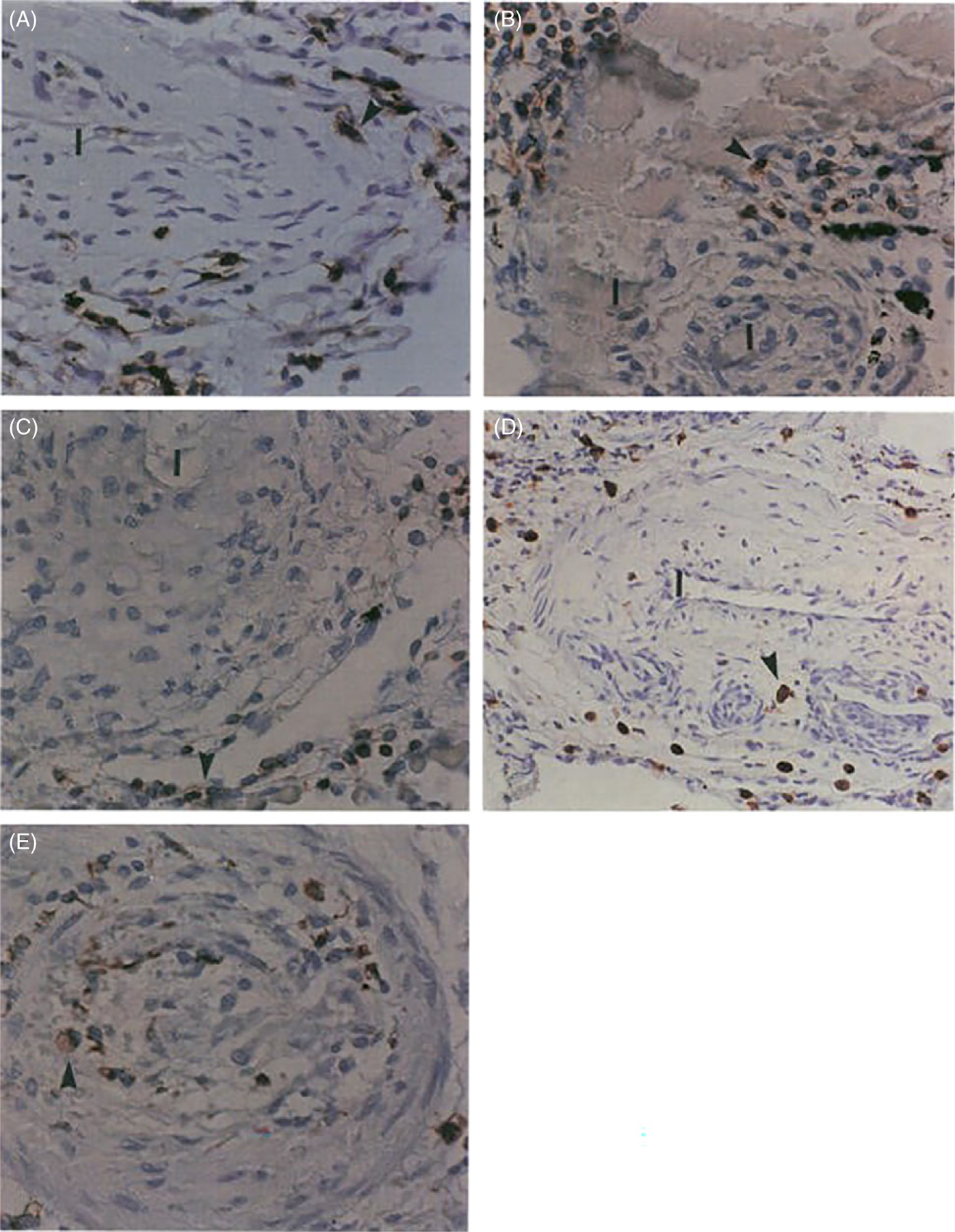
Figure 3.

Inflammatory components in plexiform type lesions of PAH. Inflammatory cells are highlighted by the anti-CD45 immunostaining (arrowheads) (A), B-cell marker (UL-26) (B), T-cell marker (UCHSC) (C), and macrophage marker (CD68) (D and E) (arrowheads). Note infiltration of the outer portion of the plexogenic vessel (D) and vessel obliterated by concentric proliferation (E) by macrophages (immunoperoxidase, A-C, E 500X; D: 200x). I-Vascular lumen. Reused, with permission, from Tuder RM, et al., 1994 (199), from Elsevier.
