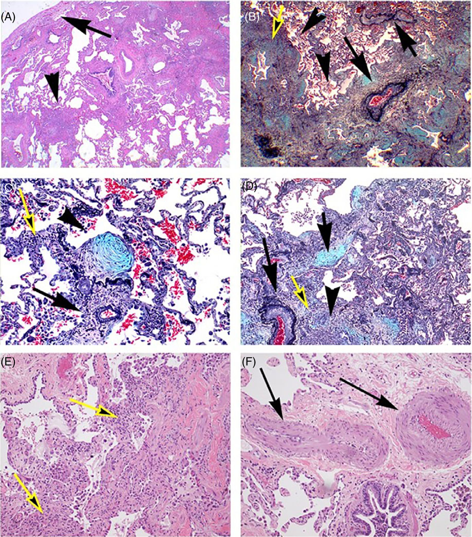
Figure 4.

Inflammatory and fibrotic processes in usual interstitial pneumonia with marked pulmonary vascular remodeling. (A) Low magnification of subpleural region showing marked subpleural fibrosis (arrow), extending irregularly toward central regions of the lung with conglomerates of lymphoid cells (arrowhead) (Hematoxilin and Eosin). (B) Marked perivascular (arrows) and airway (yellow arrow) fibrosis and inflammation, forming a continuous process along the bronchovascular and parencymal structures. The pulmonary arteries show intima thickening, with significant obliteration of the vascular lumina (Pentachrome). (C) Exudative fibroblastic focus (arrowhead) next to small pulmonary artery (arrowhead), accompanied by scattered inflammatory cells (Pentachrome). (D) Active bridging fibrosis along alveolar and bronchovascular (yellow arrow) associated with exudative process (arrow) and inflammation (arrowhead). Note the marked obliteration of an adjacent pulmonary artery (long arrow) (Pentachrome). (E) Active inflammation and interstitial fibrosis in the advancing edge of usual interstitial pneumonia (yellow arrow) (Hematoxilin and eosin). (F) Marked media and intima thickening in muscular artery (arrows) in a region not immediately affected by UIP (arrows) (Hematoxilin and eosin).
