Figure 1.

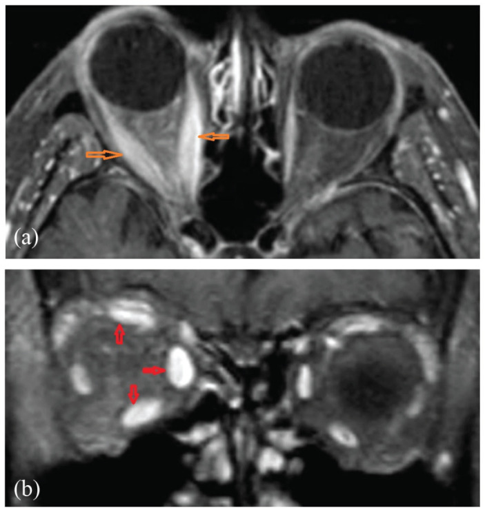
(a) Orbital axial and (b) orbital coronal; post-contrast MRI showing unilateral EOM enlargement and enhancement with tendon sparing and relative right proptosis.
MRI: magnetic resonance imaging; EOM: extraocular muscle.

(a) Orbital axial and (b) orbital coronal; post-contrast MRI showing unilateral EOM enlargement and enhancement with tendon sparing and relative right proptosis.
MRI: magnetic resonance imaging; EOM: extraocular muscle.