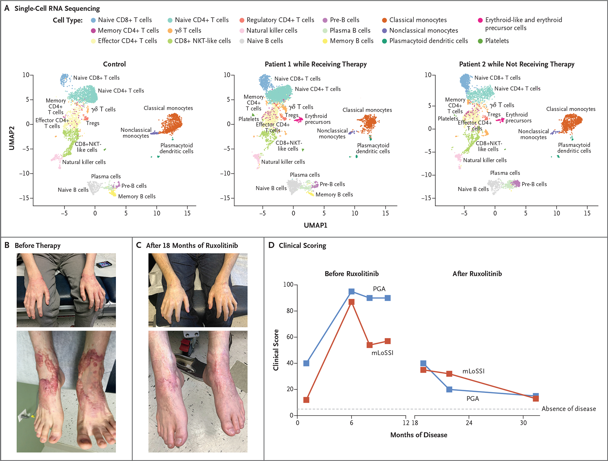
Figure 5. Ruxolitinib Treatment and Resolution of the Inflammatory Phenotype.

Shown are peripheral-blood mononuclear cells analyzed by means of single-cell RNA sequencing (Panel A), plotted in uniform manifold approximation and projection (UMAP) space and clustered for each of the control and patient samples. NKT cells denote natural killer T cells, and Tregs regulatory T cells. Photographs show waxy, erythematous nodular lesions on bilateral hands and feet before (Panel B) and after (Panel C) initiation of ruxolitinib therapy. Shown are clinical scores (Panel D) on the modified Localized Scleroderma Skin Severity Index (mLoSSI, red) and the Physician Global Assessment (PGA, blue) of disease activity. Scores on both scales range from 0 to 100, with higher scores indicating greater disease activity.
