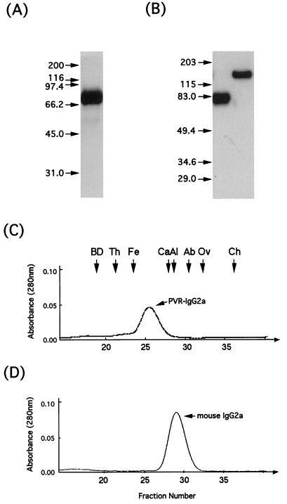
FIG. 3.
Purified recombinant PVR-IgG2a. Purified recombinant PVR-IgG2a was analyzed by polyacrylamide gel electrophoresis followed by silver staining (A) or Western blotting (B) to detect the protein, as described in Materials and Methods. Purified recombinant PVR-IgG2a (C) and, as a control, mouse IgG2a (D) were also analyzed by gel filtration. For Western blot analysis (B), PVR-IgG2a was examined under reducing (left lane) and nonreducing (right lane) conditions. Positions of molecular mass markers (in kilodaltons) are indicated by arrows on the left of panels A and B and at the top of panel C. BD, blue dextran 2000; Th, thyroglobulin (669 kDa); Fe, ferritin (440 kDa); Ca, catalase (232 kDa); Al, aldolase (158 kDa); Ab, albumin (67 kDa); Ov, ovalbumin (43 kDa); Ch, chymotrypsinogen A (25 kDa).

