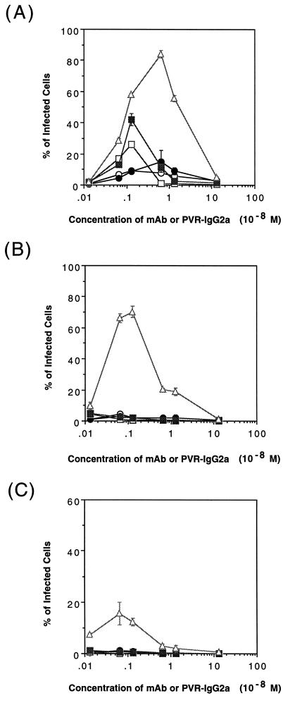
FIG. 5.
Infectivity of PV1 mediated by the anti-PV1 MAbs or PVR-IgG2a. LmFcRI cells (4.0 × 10⁴) were challenged with PV1 in the presence of anti-PV1 MAbs (7m012 [open circles], 7m039 [closed circles], Mah45i [open squares], and Mah49e [closed squares]) or PVR-IgG2a (open triangles) at the indicated concentrations. The concentrations of PV1 used were 1.3 × 10⁻¹⁰ M (9.6 × 10⁹ virions), 1.3 × 10⁻¹¹ M (9.6 × 10⁸ virions), and 1.3 × 10⁻¹² M (9.6 × 10⁷ virions) (A, B, and C, respectively). At 8 h p.i., indirect immunofluorescence was performed to detect viral antigens. The data represent means from three independent experiments, and error bars indicate standard deviations.

