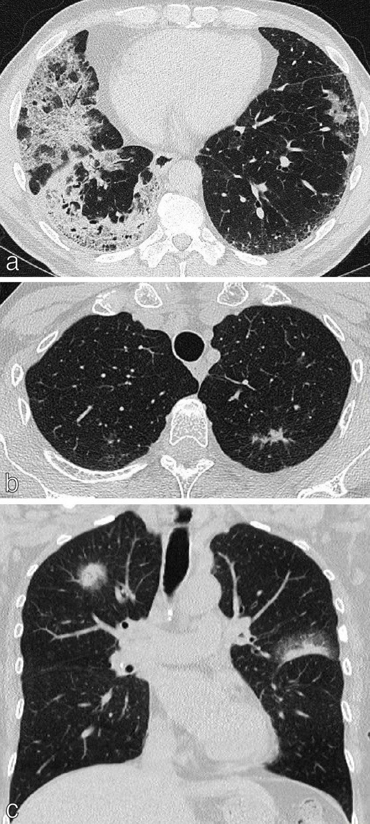
Figure 3.

Lung cancer with multiple sites of involvement. (a) Axial CT chest of a 54-year-old ex-smoker presenting with bronchorrhea showing multiple areas of consolidation and ground-glass opacification; histologically confirmed as mucinous adenocarcinoma. (b) Axial CT chest of a 60-year-old female showing a 1.5 cm left upper lobe spiculate nodule histologically confirmed as NSCLC. Multiple ground-glass nodules bilaterally represent AIS/MIA. (c) Coronal CT chest of a 66-year-old female showing solid right upper lobe and left upper lobe tumours with peripheral ground-glass (largest 2.5 cm) plus multiple ground-glass opacities in the lungs; histologically proven as non-mucinous adenocarcinoma. These were staged as multiple synchronous primaries, T1C (multiple) disease. AIS, adenocarcinoma in situ; MIA, minimally invasive adenocarcinoma; NSCLC, non-small cell lung cancer.
