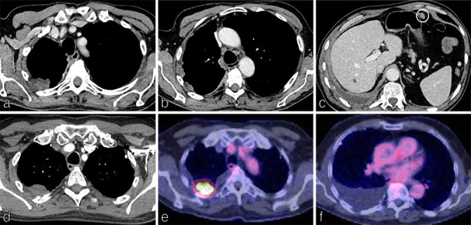
Figure 6.

Staging malignant pleural mesothelioma (MPM). Axial CT images of the chest and upper abdomen on soft tissue windows in an 83y man presenting with chest pain shows irregular pleural thickening involving the right pleura (a) and including the mediastinal surface (b) with associated volume loss and a shallow right pleural effusion (c). A metastatic deposit is identified anterior to the stomach (c) making this stage IV disease; confirmed as epitheloid MPM on histology. Axial CT chest on soft tissue windows (d) and corresponding 18F-FDG PET/CT (e, f) demonstrate a focus of irregular soft tissue in the pleura of the right upper lobe with invasion into the adjacent posterior chest wall, rib destruction plus a right pleural effusion, T4 tumour.
