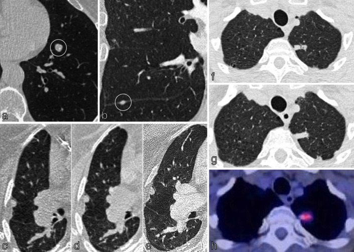
Figure 7.
Identification and follow up of pulmonary nodules. In both the BTS and Fleischner guidelines, nodules with clear features of benignity are not followed up. (a) Axial chest CT of a 66y smoker shows a left lower lobe nodule with calcification and central fat attenuation typical for a hamartoma; no follow up. (b) Coronal chest CT in a 60y smoker shows a lentiform/triangular shaped nodule associated with the horizontal fissure, typical for intrapulmonary lymph node; no follow up. The risk of malignancy in pulmonary nodules can be calculated using the Brock risk prediction tool. Nodules with < 10 % risk malignancy which are 80 mm3 (6mm) are followed up at three months and one year from baseline. (c-e) Axial CT chest imaging of an incidentally detected 9mm right upper lobe solid pulmonary nodule at baseline (c), 3 months (d) and one year (e) demonstrates stability based on the 2D non-automated diameter; plan for follow up 2 years from baseline as assessed under the BTS guidelines. Nodules showing clear growth ( 25 % volume increase/volume doubling time (VDT) of < 400 days) undergo further diagnostic work-up with 18F-FDG PET/CT /biopsy. (f-h) A 17mm left upper lobe nodule assessed at baseline (f) and at year 1 (g). Clear growth was demonstrated with more than 25 % increase in volume. The SUVmax on 18F-FDG PET/CT was 2.83 (moderate) (h) with a Herder score of 89.1 % and subsequent plan for image-guided biopsy and MDT discussion regarding treatment options.

