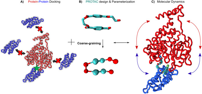
Fig. 2.
Important steps in PROTAC design for drug discovery campaigns. (a) Protein–protein docking either at the atomistic (ribbons) or coarse-grained level (red and cyan spheres). The E3 ligase is represented in red and the target protein in blue. (b) Coarse-graining of a small -molecule using the Martini 3 force field. (c) Dynamical motions of the ligase and the target (blue and red arrows, respectively) are important to query ternary complex stability in the presence of the PROTAC (represented as van der Waals spheres). All figures were rendered using VMD (Humphrey et al., 1996). The ternary complex structure is from Nowak et al. (2018) with the PDB ID code 6BN7.

