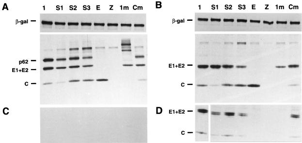
FIG. 3.
SDS-PAGE analysis of SFV structural proteins in cells cotransfected with the SFV-lacZ replicon and different SFV-helper RNAs. At 12 h posttransfection, cells were pulse-labeled for 15 min and chased for 5 min (A and C) or 3 h (B and D). Cell lysates were immunoprecipitated with a β-Gal-specific MAb (A and B, upper panels) or with a cocktail of MAbs specific for the SFV structural proteins (capsid, E2 and E1) (A and B, lower panels). Virus was sedimented from supernatants as described in Materials and Methods (C and D). SFV-lacZ RNA was coelectroporated with the following: lane 1, SFV-helper-1; lane S1, SFV-helper-S1 plus SFV-helper-C; lane S2, SFV-helper-S2 plus SFV-helper-C; lane S3, SFV-helper-S3 plus SFV-helper-C; lane E, SFV-helper-E plus SFV-helper-C; lane Z, no RNA; lane 1m, SFV-helper-1-S219A; lane Cm, SFV-helper-S-1 plus SFV-helper-C-S219A. The upper portions of panels A and B were exposed only one-third of the time compared to the lower panels; in panel D, lane 1 was exposed one-tenth of the time compared to the other lanes. The capsid protein of SFV often smears in SDS-PAGE and is not a good indicator of quantitation.

