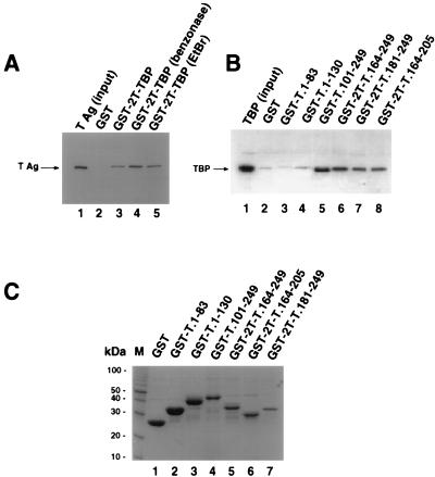
FIG. 1.
TBP binds within the DNA binding domain of T antigen. (A) Full-length T antigen (TAg; 0.5 μg) was incubated with 10 μl of glutathione-agarose containing GST (lane 2) or GST-TBP (lanes 3 to 5), either in buffer alone (lane 3) or in the presence of 1 U of benzonase (lane 4) or 200 μg of ethidium bromide (EtBr) per ml (lane 5). After washing, the beads were boiled in sample buffer, and the eluted proteins resolved by SDS-PAGE (12.5% polyacrylamide gel). T antigen was detected by immunoblotting using the T-antigen-specific monoclonal antibody Pab419. Lane 1 shows 0.1 μg of the input T antigen. (B) Full-length TBP was released from purified GST-2T-TBP by thrombin cleavage; 0.5 μg of cleaved TBP was incubated with 10 μl of glutathione-agarose containing GST (lane 2) or the indicated GST-T antigen fusion proteins in the presence of 1 U benzonase (lanes 3 to 8). After the beads were washed, the proteins were eluted by boiling in sample buffer and analyzed by SDS-PAGE (12.5% polyacrylamide gel). TBP was detected by immunoblotting using the monoclonal anti-TBP antibody 4C8. As a control, 0.1 μg of the input TBP was analyzed in parallel (lane 1). (C) Beads (10 μl) containing the GST fusion proteins used for panel B were analyzed by SDS-PAGE (12.5% polyacrylamide gel) and staining with Coomassie brilliant blue. M, 10-kDa marker protein ladder.

